Cooper ligament tear: a case - report
Abstract
We report a 34 years-old woman who presented a Cooper’s ligament tear after sudden chest torsion. This condition is extremely rare (to our knowledge, this is the first report published in the literature) but its inclusion in the differential diagnosis of traumatic chest conditions must be considered, especially since the woman is increasing the participation in sport in the last decade.
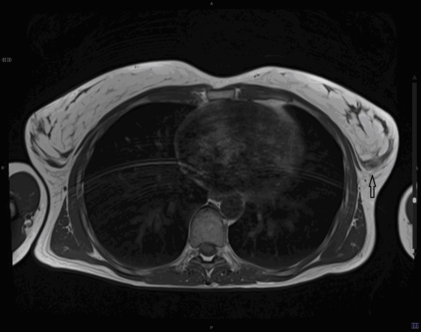
Figure 1. Thoracic magnetic resonance, axial SET1 sequence. An image of a heterogeneous signal in posterior recess of the left mammary gland with loss of contours and thickening with respect to the contralateral breast, compatible with Cooper's ligament injury is evident (arrow).

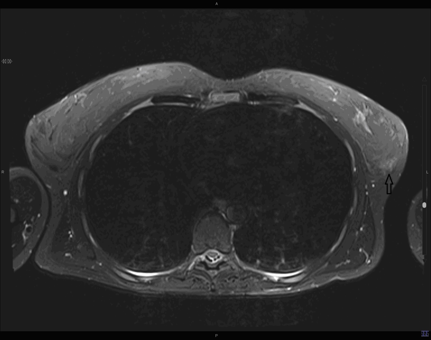
Figure 2.- Thoracic magnetic resonance, axial STIR sequence at the same cutting height as figure 1. Focus of edema located in the same area in the posterior recess of the left mammary gland, without observing the continuous ligamentous band, compatible with traumatic injury Cooper's acute ligament (arrow).


